Swanson’s Family Medicine Review 2017
Ch. 103 – Diaper Rash and Other Infant Dermatitis
A 2-Month-Old Infant with a Rash on His Cheeks
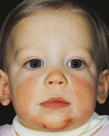
A 2-month-old infant is brought to your office by his mother. He developed an erythematous, dry rash on both cheeks approximately 1 week ago. Although the rash is always present, the mother states that it seems to be worse after she feeds him.
The mother breast-fed for the first 4 weeks of life, but she returned to work 4 weeks ago and switched the baby from breastfeeding to bottle-feeding. The mother has a history of asthma, and the father has seasonal allergies.
On examination, the child appears healthy. He has an erythematous maculopapular eruption that covers his cheeks, and he appears to be developing an erythematous rash on his neck, both wrists, and both hands (Fig. 103-1). The rest of the physical examination is within normal limits.

- What is the most likely cause of this infant’s rash? atopic dermatitis
Atopic dermatitis is an inflammatory skin disease characterized by erythema, edema, pruritus, exudation, crusting, and it is often referred to as the “itch that rashes.” Atopic dermatitis can be classified as infantile (up to 2 years), childhood (2 to 12 years), and adult atopic dermatitis. Infantile atopic dermatitis usually is manifested at 4 months of age, and 50% of cases resolve by 18 months. The areas most commonly affected include the cheeks, neck, wrists, hands, and extensor aspects of the extremities. Spread often occurs from extensor to flexor surfaces. Pruritus may lead to intense scratching and secondary infection. In older children, flexural surfaces are involved, often with xerosis and lichenification. Atopic dermatitis may be exacerbated by food allergies (in approximately 40% of cases), such as from eggs, wheat, peanuts, or cow’s milk; environmental stimuli, such as dust mites or animal dander; or emotional stress.
- What is (are) the recommended initial treatment(s) of the rash in the infant presented?
- skin hydration with petroleum products
- local corticosteroid therapy
- minimizing bathing and the use of soap
The treatment of atopic dermatitis begins with the avoidance of any environmental factors that precipitate the condition and also maintenance of the skin barrier. Disruption of the acid mantle of the skin by alkaline soaps is a major factor in the development and worsening of the condition. The most important maintenance therapy is to use non-soap alternatives (Aveeno, Cetaphil, and Dove) or pH-neutral cleaners and to bathe only dirty skin areas. In addition, emollients (e.g., petroleum products) to restore skin hydration should be applied within 3 minutes of bathing. Excessive drying of the skin should be avoided. Atopic dermatitis is best managed with local therapy. Flare-ups of the condition are treated with topical corticosteroid creams or lotions. To further prevent scratching, the fingernails should be cut short. Percutaneous absorption of corticosteroid does occur, and atrophy of the skin should be watched for. This can be avoided by use of only lower moderate-potency topical corticosteroids. Topical calcineurin inhibitors such as pimecrolimus (Elidel) are not approved for infants younger than 2 years, especially since the addition of the United States Food and Drug Administration (FDA) black box warning for lymphomas and skin cancer, but they are used for second-line therapy in older children and are still used by some physicians in place of high-potency steroids in younger children with severe atopic dermatitis. Systemic antihistamines and nonsedating antihistamines have to be used to control pruritus, but not in infants. These can improve sleep and prevent more excoriations and secondary infections. Secondarily infected atopic dermatitis may often be managed by topical mupirocin, but it may occasionally require systemic treatment.
- Which statement is true regarding the prognosis and treatment of this condition?
- 50% of these patients will improve by 18 months
- because topical corticosteroids rarely control symptoms, systemic steroids are usually required
- up to 80% of these patients will develop allergy or allergic rhinitis symptoms
Atopic dermatitis is largely a condition of children. Nearly 60% present with symptoms as infants in the first 12 months of life, and another 25% of patients will have disease presentation by 5 years of age. Of those who present in infancy, 50% will resolve by 18 months. Topical corticosteroids and treatment with skin hydration are adequate therapy for most infants and young children, but many also require treatment with topical calcineurin inhibitors (older than 2 years) and antihistamines.
A 3-Week-Old Infant with a Crusty Head
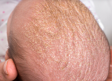
A grandmother brings in a 3-week-old infant with the complaint that she has “crusting” on her hair. Because it has been quite cold, the grandmother (who cares for the child) has been careful not to bathe the child too often, and she does not use shampoo. The child is bottle-feeding and has otherwise had no health problems. On examination, the infant appears healthy and interactive. Her scalp is covered with a crusty, yellowish rash with some erythema and crusting at the base of the ears and scalp (Fig. 103-2).

- The most likely diagnosis of this rash is: seborrheic dermatitis
Pityriasis capita or cradle cap (diffuse or focal scaling and crusting of the scalp) is a common form of seborrheic dermatitis seen in infancy. Seborrheic dermatitis is most common in children during the first 3 months of life. Typically, it is a dry, scaly, erythematous, papular dermatitis that is usually nonpruritic. It may involve the face, neck, retroauricular areas, axillae, and diaper area. The dermatitis may be patchy or focal, or it may spread to involve the entire body. Although it is common, the etiology of seborrheic dermatitis is unclear, but it is possibly related to the proliferation of the Malassezia species. Hormonal and genetic factors as well as alteration of essential fatty acid patterns are also thought to play a major role.
- Recommended treatment of the rash initially is: gentle cleaning of the scalp with a soft washcloth and baby shampoo, using oil to remove scales if needed
In general, gentle cleaning with a soft washcloth, occasionally using baby oil or mineral oil to remove scales, is curative for cradle cap. If the seborrheic dermatitis extends onto the face or persists, a low-dose hydrocortisone cream may be used for a short time or selenium shampoo may be used on the scalp. Caretakers are often afraid to shampoo the hair, causing buildup of the crust
A 2-Week-Old Infant Who Looks “Too Ugly for Photos”
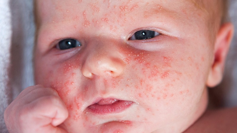
A 2-week-old infant, a product of a normal birth and delivery, is brought to your office by his mother, who says she cannot take baby photos because his skin “won’t clear up.” According to the mother, ever since birth, the child has had small pimple-like lesions on his face and scattered over his body that come and go (Fig. 103-3). They sometimes get slightly erythematous, but they are not crusty. The baby is not bothered by them, but his face looks “so ugly” that his mother does not want to have his photo taken. She wants medicine to “get rid of it.”

- What is the most likely diagnosis in this infant? infantile acne
This infant has infantile acne, a benign condition that generally presents problems only of appearances and for It spontaneously resolves and does not require treatment. It should be distinguished from atopic dermatitis, seborrheic dermatitis, erythema toxicum neonatorum, and neonatal pustular melanosis. Erythema toxicum neonatorum usually appears soon after birth and lasts up to 2 weeks. It is characterized by vesicles, pustules, and papules on an erythematous halo base. Transient pustular neonatal melanosis, more common in dark-skinned infants, also has vesicles and pustules but no erythematous base, and it may have a collarette of scale. Milia are epidermal inclusion cysts, and miliaria is essentially a “heat rash.”
- What is (are) the treatment(s) of choice for the condition described here?
Reassurance that it will clear, with an understanding that no treatment is necessary, is important. None of the other treatments listed is necessary.
An 8-Month-Old Infant with a Long-Term Persistent Diaper Rash
An 8-month-old infant is brought to your office by his mother for assessment of a diaper rash. His mother has tried cornstarch powder, vitamin E cream, zinc oxide, and a corticosteroid cream prescribed by a different physician.
On examination, the infant has an intensely erythematous diaper dermatitis that has a scalloped border and a sharply demarcated edge. Numerous satellite lesions are present on the lower abdomen and thighs (Fig. 103-4).

- What is the most likely diagnosis in this infant? candidal diaper dermatitis
This infant has candidal diaper dermatitis, which is manifested as an erythematous confluent plaque formed by papules and vesiculopustules, with a scalloped border and a sharply demarcated. Candidal diaper dermatitis can usually be distinguished from other childhood diaper dermatoses by the presence of satellite lesions produced at some distance from the primary eruption. Although this child’s rash is not primarily perianal, streptococcal dermatitis should be considered in the case of a chronic perianal rash. Diagnosis can be made by culture, and it responds to treatment with amoxicillin. Intertrigo, common in infants with overlapping skin and fat folds, usually responds to treatment to decrease the moisture. Primary irritant dermatitis is discussed later.
- What is the treatment of choice for this infant’s diaper rash? a topical antifungal agent
The treatment of choice in candidal diaper dermatitis is a topical antifungal agent. Topical miconazole, clotrimazole, or ketoconazole is commonly used. In an infant with a severe inflammatory reaction, a topical corticosteroid may be mixed 50/50 with a topical antifungal agent and applied on a regular basis for a few days to 1 week. Rather than a high-potency steroid–antifungal combination, however, a low-potency steroid such as hydrocortisone with the antifungal agent is preferred because the infant is at risk for striae and local complications of steroid therapy.
A 4-Month-Old Infant with a Diaper Rash Caused by Dirty Diapers
A 4-month-old infant is brought to your office by her mother. Her mother complains that the child has a diaper rash that is probably related to her “not changing the diapers often.” The mother has five other children and was trying to save money by using fewer diapers. She admits that she waits until the diaper is soaked before changing it.
On examination, the infant has erythematous, scaly, papulovesicular diaper dermatitis with numerous bullous lesions, fissures, and erosions.
- What is the most likely diagnosis in this infant? primary irritant contact dermatitis
This child has a primary irritant contact dermatitis. Irritant contact dermatitis is a reaction to friction, maceration, and prolonged contact with urine and feces. It usually presents as an erythematous, scaly dermatitis with papulovesicular or bullous lesions, fissures, and erosions. The eruption can be either patchy or confluent. The genitocrural folds are often spared. Secondary infection with either bacteria or yeast can occur. The infant can be in considerable discomfort because of the marked inflammation that is sometimes associated with this type of diaper rash. Primary irritant diaper dermatitis should be managed by frequent changing of diapers and thorough washing of the genitalia with warm water and a mild soap. Occlusive plastic pants that promote maceration should be avoided. If cloth diapers are used, they should be changed frequently, as should disposable diapers. Drying out the skin by allowing the infant to go without diapers can be helpful but can be logistically challenging. An occlusive topical agent such as zinc oxide or A&D ointment can be applied until healing occurs.
- What differentiates a candidal diaper rash from irritant diaper dermatitis? irritant diaper dermatitis spares the crural folds; candidal diaper rash has satellite lesions
A useful diagnostic pearl for differentiation of candidal diaper rash from irritant diaper dermatitis is that candidal diaper rash tends to involve the warm, moist folds of the skin, whereas irritant diaper dermatitis tends to spare the folds, occurring mainly in the areas of greatest skin contact with urine and feces. Candidal diaper rash also tends to present with satellite lesions and will worsen with prolonged topical corticosteroid therapy.
Summary of Skin Problems
Atopic Dermatitis
It usually begins and is more prominent on the cheeks of infants and treatment includes: avoidance of soaps and limited bathing frequency; use of agents to promote skin hydration; low-dose corticosteroids may be used, and in children older than 2 years or in particularly severe cases in younger children, topical calcineurin inhibitors may be used (Elidel is approved for children older than 2 years). There is a strong association with allergies and asthma; 80% of patients develop these, and there is often a strong maternal family history of these illnesses.
Seborrheic Dermatitis
Cradle cap typically presents in early infancy and can be treated by gentle cleaning with soap and removal of scales with oil; low-dose corticosteroids and occasionally antifungal agents may be used if needed.
Candidal Dermatitis
A diagnostic clue is the satellite lesions around the peripheral area of the main area of dermatitis.
Treatment consists of topical miconazole or topical clotrimazole. A mild topical hydrocortisone mixed with a topical antifungal agent can be used when severe inflammation is present.
Primary Irritant Dermatitis
This rash presents with maceration, often a history of infrequent diaper changes, and prolonged exposure to urine and feces. It is treated with an occlusive topical agent such as zinc oxide or petroleum jelly or zinc oxide applied over hydrocortisone base when severe inflammation is present; frequent diaper changes are essential. Erythema toxicum neonatorum, milia, miliaria, and transient pustular neonatal melanosis are self-limited skin conditions of the newborn that do not require treatment.
- Which of the following is not routinely associated with atopic dermatitis? Immunodeficiency
Atopic dermatitis is often associated with a family history of allergies, asthma, hay fever, or atopic dermatitis. Individuals are more sensitive to certain fibers, particularly wool. Rough fibers tend to be more irritating than smooth fibers in clothing textiles. Keratosis pilaris, which is characterized by asymptomatic horny follicular papules on the upper arms, buttocks, and thighs, is another manifestation of atopic dermatitis. Immunodeficiency (e.g., severe combined immunodeficiency, Wiskott-Aldrich syndrome) should be considered in an infant with a rash that does not resolve, but it is not typical of atopic dermatitis in general.
- Which statement is true regarding the etiology of atopic dermatitis?
- A maternal history of allergy or asthma is the most important genetic predictor
- Breastfeeding offers advantage over formula feeding in this condition
- Eosinophilia is common in these patients
- There is an increased production of allergen-specific T cells and some interleukins, leading to elevated immunoglobulin E levels in patients with this condition
- Food allergies are important triggers in 40% of young children with this condition
Breastfeeding is important to prevent atopic dermatitis, particularly if breastfeeding is exclusive and continued until the infant is older than 6 months.
- For primary irritant contact dermatitis, what is the treatment of choice for an infant? Zinc oxide paste or other barrier ointments
An occlusive topical agent such as zinc oxide or A&D ointment can be applied until healing occurs. Topical 1% hydrocortisone ointment is also useful in the management of diaper dermatitis in its more severe form. Neither systemic antibiotics nor systemic corticosteroids are indicated in the treatment of primary irritant diaper dermatitis. Again, the primary treatment is frequent diaper changing or leaving the child without a diaper as much as possible until the skin heals.
Suggested Reading
Behrman RE, Kliegman R, Jenson H. Nelson textbook of pediatrics. ed 17. Philadelphia: Saunders; 2004.
Boiko S. Making rash decisions in the diaper area. Pediatr Ann. 2000;29:50–56.
Habif TB. Clinical dermatology: a color guide to diagnosis and therapy. ed. 4 St. Louis: Mosby; 2004.
Scheinfeld N. Diaper dermatitis: a review and brief survey of eruptions of the diaper area. Am J Clin Dermatol. 2005;6:273–281.


